 IOL-Experience
 Technologies
 IOL calculators
 Service & handling
 Sustainability
 Congresses and events
 Download Center
How safe is the implantation of a 1stQ AddOn Sulcus IOL?
10.09.2026 (updated: 21.09.2026)Sulcus-fixated 1stQ AddOn intraocular lenses offer the option of correcting refractive errors or extending the range of vision without exchanging the primary capsular bag lens. This article summarises the published evidence on safety, stability, intraocular pressure (IOP), pigment dispersion, interlenticular opacification and reversibility.
Summary
Sulcus-fixated AddOn intraocular lenses enable secondary refractive correction or a subsequent extension of the range of vision without the need to exchange the IOL originally implanted in the capsular bag. Published data on the 1stQ AddOn cover refractive, toric, EDOF and trifocal applications, as well as the primary dual lens procedure Liberty².
Across the available clinical studies, a total of 217 eyes received sulcus-fixated supplementary lenses that are comparable in terms of their haptics, shape and basic sulcus-specific design. In vitro and anatomical investigations provide additional evidence. The studies differ in design, indication, follow-up duration and patient selection and should therefore not be interpreted as a formal pooled analysis.
Taken together, however, they support a consistent picture: stable sulcus positioning, no persistent IOP elevations, no clinically relevant signs of pigment dispersion and no interlenticular opacification were reported in the cohorts studied. [1–10]
An additional safety aspect of the concept is reversibility: where clinically indicated, the sulcus-fixated supplementary lens can be removed or exchanged while retaining the primary capsular bag lens. This was demonstrated in clinical practice in a published case report describing the bilateral exchange of sulcus-fixated trifocal supplementary lenses. [7]
Why does the safety of a sulcus IOL require particular consideration?
An additional IOL in the ciliary sulcus is anatomically closer to the iris and ciliary body than a conventional capsular bag lens. Three safety aspects are therefore particularly relevant to sulcus IOLs:
• Stable positioning and avoidance of decentration, tilt or clinically relevant rotation.
• Avoidance of iris or uveal contact and the resulting pigment dispersion.
• Avoidance of a secondary increase in intraocular pressure.
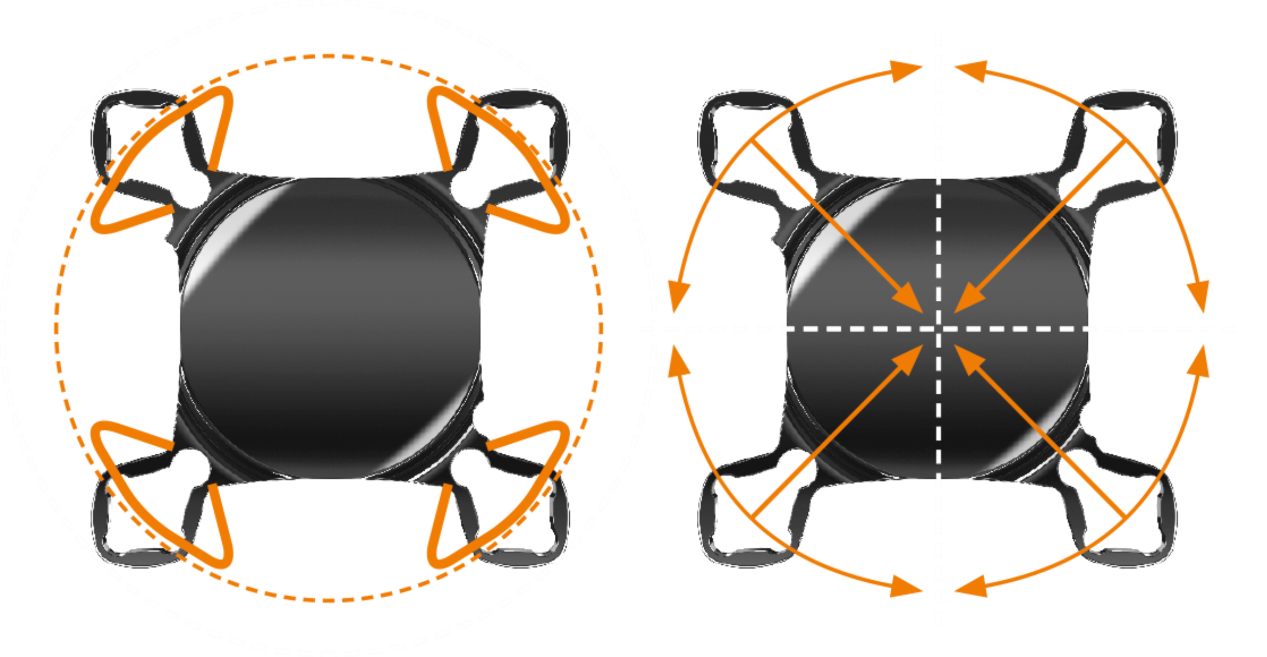
With two lenses in the eye, the possibility of interlenticular opacification must also be considered. Published studies and technical investigations therefore emphasise that a supplementary IOL must be specifically designed for placement in the sulcus. The 1stQ AddOn has a dedicated design featuring four flexible haptics, an overall diameter of 13 mm, a 6 mm optic, a square design and a convex–concave optic.
These features address sulcus positioning, separation from the capsular bag lens, rotational stability and reduced iris interaction. [2,6,9,10]
| Design feature | Safety-related objective |
|---|---|
| Four flexible haptics | Adaptation to the sulcus and support for rotational stability |
| Square design | Addresses iris capture |
| Convex–concave optic | Separation from the capsular bag lens; reduction of the risk of interlenticular opacification |
| Hydrophilic acrylic | Biocompatible material design intended for sulcus placement |
| 6 mm optic | Optical function with stable sulcus positioning |
What do the clinical safety data show?
2.1 Intraocular pressure
Postoperative intraocular pressure is one of the most important safety parameters in sulcus implantation. In the toric study by Gundersen and Potvin involving 18 eyes, mean IOP remained virtually unchanged before and after secondary implantation: 12.6 ± 2.6 mmHg before implantation versus 12.9 ± 2.4 mmHg at follow-up; no eye had an IOP above 20 mmHg. No pigment dispersion or interlenticular opacification was observed. [2]
Early postoperative IOP elevations also occurred in the study of the primary dual lens procedure Liberty² by Harrisberg et al., which included 32 eyes in the dual lens procedure group and 57 eyes in the comparison group. However, these elevations occurred in both groups. At one year, IOP values were within the normal range in both groups, with no significant differences between them. [6]
Interpretation: The available data provide no indication of a clinically relevant chronic IOP elevation associated with the sulcus-specific 1stQ AddOn in the cohorts studied. Nevertheless, early postoperative IOP elevations remain an important consideration in perioperative care. [1,2,4,6]
2.2 Pigment dispersion and iris interaction
A key potential complication of a sulcus IOL is mechanical contact with the iris. With unsuitable IOL designs, this may lead to pigment dispersion, iris defects and secondary IOP elevations. No corresponding pattern of complications was reported for the 1stQ AddOn in the available clinical studies.
The toric study involving 18 eyes found no evidence of pigment dispersion. Clinical studies of the primary dual lens procedure likewise found no associated persistent IOP elevation or corresponding pattern of complications. The available data therefore provide a consistent picture of good uveal tolerability. However, this statement is limited to the cohorts and follow-up periods studied. [2,3,4,6]
How stable is the AddOn in the sulcus?
Mechanical stability is particularly relevant to toric AddOn lenses because rotation directly affects the effectiveness of cylindrical correction. In the study by Gundersen and Potvin involving 18 eyes, the mean absolute deviation from the intended axis was 4.9 ± 3.7°. Sixteen of the 18 eyes (89%) were within 10° of the intended axis; no eye showed a deviation greater than 13°. Repositioning was not required. [2]
The earlier study by Gundersen and Potvin on secondary AddOn implantation also provides clinical data from 46 eyes; a significant reduction in refractive astigmatism was reported in the toric cases. [1,2]
Of relevance to clinical safety: Stable sulcus positioning reduces both the risk of refractive miscorrection and the likelihood of optically significant decentration. Anatomical investigations in pseudophakic cadaver eyes further support the feasibility of suitable positioning of the hydrophilic supplementary IOL studied for sulcus fixation. However, these data are preclinical and should not be equated with long-term clinical evidence. [10]
Of relevance to clinical safety: Stable positioning reduces both the risk of refractive miscorrection and the likelihood of optically significant decentration. [1,9]
Is there evidence of interlenticular opacification?
Historical piggyback approaches involving two lenses in the capsular bag have been associated with interlenticular opacification. The 1stQ AddOn follows a different approach: the second lens is positioned in the sulcus and features a convex–concave optic designed to maintain separation from the capsular bag lens. [2,9,10]
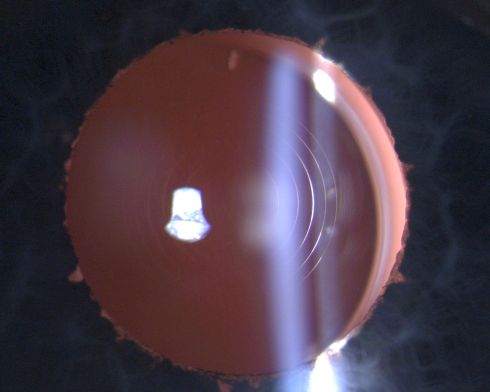
In the toric clinical study involving 18 eyes, no interlenticular opacification was observed in any eye. Similarly, the study of the primary dual lens procedure by Harrisberg et al. reported neither IOL opacification nor interlenticular opacification during the one-year follow-up. [2,6]
The available data therefore provide no evidence of interlenticular opacification in the cohorts studied. However, the existing studies do not allow reliable conclusions about the frequency of very rare opacification events or those occurring after longer periods.
What is known about the cornea and endothelium?
Endothelial tolerability is another relevant safety parameter when implanting an additional intraocular lens. In the study by Palomino-Bautista et al., 18 eyes of 11 pseudophakic patients were followed for six months. In addition to visual acuity and patient satisfaction, assessments included IOP, biomicroscopic findings and endothelial cell count; no adverse changes in endothelial cell count were reported. [3]
Limitation of the evidence: In the study by Harrisberg et al., endothelial cell count was not systematically evaluated as a primary safety parameter. However, the authors reported no corneal decompensation in the dual lens procedure group during the one-year follow-up. [6]
Interpretation: There are favourable indications of endothelial and corneal tolerability. However, the data remain limited in terms of sample size, follow-up duration and systematic long-term endothelial measurements. Cautious wording is therefore recommended for the website article: no adverse endothelial findings were reported in the available investigations, but comprehensive long-term evidence spanning many years is lacking. [3,6]
What happens if the AddOn is not tolerated?
A key difference compared with a multifocal capsular bag IOL is the reversibility, in principle, of the sulcus-fixated supplementary lens. The AddOn is located in the sulcus and can be removed or exchanged where clinically indicated, while preserving the refractive function of the primary capsular bag lens. [6,7,9]
In a published case report, the sulcus-fixated supplementary IOL in each eye was exchanged six months after a bilateral dual lens procedure because of unacceptable residual refractive error and photic phenomena. The procedures were described as uneventful; three months later, visual acuity was −0.22 and −0.20 logMAR, respectively, and endothelial cell counts remained stable. [7]
Interpretation: Reversibility is therefore not merely a theoretical concept; it has been clinically documented. However, this evidence comes from a case report and cannot be used to derive a general complication rate for explantation or exchange.
How safe is implantation during primary cataract surgery?
Harrisberg et al. compared 32 eyes undergoing the primary dual lens procedure Liberty²—a monofocal capsular bag lens plus a trifocal 1stQ AddOn in the sulcus—with 57 eyes receiving a single multifocal capsular bag lens. At one month and one year, there were no significant differences in uncorrected distance or near visual acuity. There were also no significant differences in IOP between the groups at any assessment time point. [6]
This study therefore provides a direct clinical comparison between a primary dual lens procedure and a multifocal capsular bag IOL. The results suggest that, in this selected cohort, the additional sulcus lens was not associated with an identifiable additional long-term safety disadvantage. [6]
However, several factors must be considered: the study was retrospective and conducted at a single centre, and the dual lens procedure group comprised 32 eyes. Certain patients at increased risk were also excluded, including those with glaucoma, pigment dispersion, uveitis, retinal disease, zonular laxity or anatomical abnormalities. The results therefore cannot be readily generalised to all patient populations. [6]
What role does patient selection play?
The studies do not show that every patient is equally suitable for AddOn implantation. afety is not solely a property of the lens; it results from the combination of lens design, anatomical suitability, appropriate indication, surgical technique and postoperative monitoring.
Patient selection is particularly relevant in eyes with glaucoma or pigment dispersion syndrome, uveitis, significant retinal disease, zonular laxity, anatomical abnormalities of the anterior segment or other factors that may complicate sulcus positioning or follow-up. Harrisberg et al. excluded patients with these risk factors. [6]
The study of partial monovision with a unilaterally implanted multifocal AddOn also notes that supplementary implantation constitutes an additional surgical procedure. AddOn implantation is described there as brief and atraumatic; nevertheless, every additional intraocular operation carries its own risk profile. [8]
What do the studies show overall?
| Safety parameter | Findings from the expanded evidence base | References |
|---|---|---|
| IOP | No evidence of persistent IOP elevation in the AddOn cohorts studied; early postoperative pressure elevations were observed but were not exclusive to the AddOn group. | [1,2,3,4,6] |
| Pigment dispersion | Not observed in the relevant clinical studies of the 1stQ AddOn, or no corresponding persistent pattern of complications reported. | [2,3,4,6] |
| Iris Capture / Iris interaction | Addressed by the square design and sulcus-specific haptic design; not identified as a major safety concern in the reported studies. | [2,6,9,10] |
| Interlenticular opacification | Not observed in the toric study or the one-year study of the primary dual lens procedure. | [2,6] |
| Centration / positioning | Good sulcus positioning reported clinically; preclinical anatomical data support positioning in the pseudophakic eye. | [1,2,10] |
| Toric rotation | Mean absolute axis deviation of 4.9 ± 3.7°; 89% within 10°, with no eye exceeding 13°. | [2] |
| Endothelium / cornea | No adverse changes in endothelial cell count in the available six-month study; no corneal decompensation reported in the one-year Harrisberg cohort. | [3,6] |
| Reversibility | Bilateral exchange of sulcus-fixated trifocal supplementary lenses clinically documented; the evidence is a case report. | [7] |
| Optical quality | An in vitro comparison shows comparable optical quality between the dual lens procedure and a trifocal capsular bag IOL; the two lenses did not substantially compromise optical quality in the experimental setup. | [9] |
Limitations of the evidence: The available studies predominantly involve smaller, indication-specific cohorts. The evidence includes retrospective studies, case series, in vitro and cadaver investigations, and a case report. Long-term data are not systematically available for all safety parameters. [1–12]
10. Conclusion: How safe is 1stQ AddOn implantation?
The currently available studies indicate a consistent, overall favourable safety and stability profile for the 1stQ AddOn when implanted in the sulcus as intended and with careful patient selection.
Particularly noteworthy findings include:
- Stable sulcus positioning, including good rotational stability of the toric version. [1,2,10]
- No evidence of persistent IOP elevations in the cohorts studied. [1,2,3,4,6]
- No pigment dispersion observed in the relevant clinical studies. [2,3,4,6]
- No interlenticular opacification observed in the reported AddOn cohorts. [2,6]
- No corneal decompensation reported in the one-year cohort undergoing a primary dual lens procedure. [6]
- Clinically documented reversibility through exchange of the sulcus-fixated supplementary lens. [7]
Across the studies, 217 eyes were examined following implantation of sulcus-fixated supplementary lenses that are comparable in terms of their haptics, shape and basic sulcus-specific design.
For design-related safety aspects—in particular sulcus positioning, iris interaction, pigment dispersion, iris capture, interlenticular opacification and IOP over time—the findings provide a consistent picture: no clinically relevant persistent IOP elevations, pigment dispersion, iris capture events or interlenticular opacification were reported in the cohorts studied.
References
[1] Gundersen KG, Potvin R. A review of results after implantation of a secondary intraocular lens to correct residual refractive error after cataract surgery. Clinical Ophthalmology. 2017;11:1791–1796. DOI: 10.2147/OPTH.S144675. Supports: Secondary AddOn implantation for refractive refinement; clinical data from 46 eyes.
[2] Gundersen KG, Potvin R. Refractive and Visual Outcomes After Implantation of a Secondary Toric Sulcus Intraocular Lenses. Clinical Ophthalmology. 2020;14:1337–1342. DOI: 10.2147/OPTH.S255725; PMID: 32546940. PubMed: https://pubmed.ncbi.nlm.nih.gov/32546940/. Supports: Toric 1stQ AddOn, 18 eyes, rotational stability, IOP, absence of pigment dispersion and interlenticular opacification.
[3] Palomino-Bautista C, Sánchez-Jean R, Carmona Gonzalez D, Romero Domínguez M, Castillo Gómez A. Spectacle Independence for Pseudophakic Patients – Experience with a Trifocal Supplementary Add-on Intraocular Lens. Clinical Ophthalmology. 2020;14:1043–1054. DOI: 10.2147/OPTH.S238553; PMID: 32308364. PubMed: https://pubmed.ncbi.nlm.nih.gov/32308364/. Supports: Trifocal supplementary IOL in pseudophakic patients; IOP, endothelial and biomicroscopic follow-up; 18 eyes of 11 patients.
[4] Albayrak S, Comba ÖB, Karakaya M. Visual performance and patient satisfaction following the implantation of a novel trifocal supplementary intraocular lens. European Journal of Ophthalmology. 2021;31(5):2346–2352. DOI: 10.1177/1120672120969042. Supports: Visual outcomes and patient satisfaction following implantation of a trifocal supplementary IOL; additional clinical data on trifocal AddOn use.
[5] Gundersen KG, Gjerdrum B, Potvin R. Efficacy of a Secondary Trifocal Sulcus IOL in Providing Near and Intermediate Vision in Patients with Prior Myopic Laser Vision Correction and Cataract Surgery. Clinical Ophthalmology. 2022;16:2219–2226. DOI: 10.2147/OPTH.S372925. Supports: Secondary trifocal sulcus IOL in pseudophakic patients following myopic laser vision correction and cataract surgery.
[6] Harrisberg BP, Chua AW, Chua MJ, Taher A. Comparison of Primary Duet Lens Procedures: In-The-Bag Monofocal with Sulcus Multifocal, and Standard Single Multifocal Lens for Cataract Surgery. Clinical Ophthalmology. 2023;17:273–282. DOI: 10.2147/OPTH.S396472; PMID: 36698853. PubMed: https://pubmed.ncbi.nlm.nih.gov/36698853/. Supports: Primary dual lens procedure Liberty², 32 eyes versus 57 control eyes, IOP over time, complications and one-year follow-up.
[7] Baur ID, Auffarth GU, Yildirim TM, Mayer CS, Khoramnia R. Reversibility of the duet procedure: Bilateral exchange of a supplementary trifocal sulcus-fixated intraocular lens for correction of a postoperative refractive error. American Journal of Ophthalmology Case Reports. 2020;20:100957. DOI: 10.1016/j.ajoc.2020.100957; PMID: 33102931. PubMed: https://pubmed.ncbi.nlm.nih.gov/33102931/. Supports: Clinically documented reversibility through bilateral exchange of sulcus-fixated trifocal supplementary lenses.
[8] Knecht VA, Colosi HA, Hassenstein A. Partial monovision achieved by unilateral implantation of a multifocal add-on lens with bilateral pseudophakia: evaluation and results. Graefe’s Archive for Clinical and Experimental Ophthalmology. 2022;260:2753–2762. DOI: 10.1007/s00417-022-05584-y; PMID: 35175409. PubMed: https://pubmed.ncbi.nlm.nih.gov/35175409/. Supports: Partial monovision with a unilateral multifocal AddOn, near and intermediate vision, spectacle independence and consideration of additional surgical risks.
[9] Khoramnia R, Baur ID, Yan W, Łabuz G, Auffarth GU. Comparison of a Presbyopia-Correcting Supplementary Intraocular Lens Combination and a Capsular-Bag Lens: An In Vitro Study. Diagnostics. 2023;13(8):1482. DOI: 10.3390/diagnostics13081482; PMID: 37189583. PubMed: https://pubmed.ncbi.nlm.nih.gov/37189583/. Supports: Optical comparability of the dual lens procedure with a trifocal capsular bag IOL in an in vitro setting.
[10] Reiter N, Werner L, Guan J, Li J, Tsaousis KT, Mamalis N, Srinivasan S. Assessment of a new hydrophilic acrylic supplementary IOL for sulcus fixation in pseudophakic cadaver eyes. Eye. 2017;31:802–809. DOI: 10.1038/eye.2016.310. Supports: Preclinical/anatomical investigation of sulcus fixation, positioning and separation in pseudophakic cadaver eyes.
[11] Hassenstein A, Niemeck F, Giannakakis K, Klemm M. Torische AddOn-Intraokularlinsen zur Korrektur hoher Astigmatismen nach pseudophaker Keratoplastik. Der Ophthalmologe. 2017;114(6):549–555. DOI: 10.1007/s00347-016-0386-6. Supports: Specific clinical application of toric AddOn IOLs for high astigmatism following pseudophakic keratoplasty.
[12] Kahraman G, Dragostinoff N, Brezna W, Amon M. Visual outcomes and patient satisfaction after bilateral sequential implantation of a capsular bag IOL and a Supplementary Sulcus-Fixated Trifocal IOL. Journal of Refractive Surgery. 2021;37(2):105–111. DOI: 10.3928/1081597X-20201215-01. Supports: Additional clinical data on sequential implantation of a capsular bag IOL and a sulcus-fixated trifocal supplementary IOL.